1. Home
  2. Specialties & Services
  3. Cartilage Preservation and Restoration
  1. Specialties & ServicesSpecialties & Services
Skip to Main Content
  • MyChart
  • Pay a Bill
  • Help
St. Luke's Logo to Homepage
Couple rowing a boat

Cartilage Preservation and Restoration

Couple rowing a boat

Cartilage Preservation and Restoration

Preserving Mobility with Advanced Cartilage Treatments

The St. Luke's cartilage preservation and restoration program is dedicated to the treatment of patients with symptomatic cartilage damage, including isolated cartilage defects, meniscal injuries and early arthritis. Our expert physicians will evaluate and recommend treatment options to match the physical demands of each patient's lifestyle.

Our philosophy is to repair the cartilage damage by treating the whole patient and determining the underlying causes of damage, which can include malalignment, ligament instability and loss of meniscal function. We'll also educate patients on the safest and latest treatment options to help them to make the most informed decision regarding their cartilage damage. Conservative, or nonsurgical, management is usually attempted before more invasive, surgical options are considered.

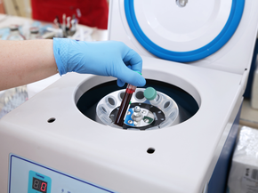
Our patients also have access to local and national cartilage repair and restoration research projects through partnerships with St. Luke's Applied Research and Boise State University.

For more information on our cartilage preservation and restoration program, please call (208) 706-9118.

woman playing pickleball

Our goal is to help you move freely and live fully so you can get back to doing what you love at home, at work, or at play.

Non-Surgical Approaches to Cartilage Repair and Preservation
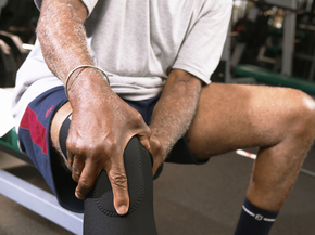
  • Man holding his knee in gym

    Lifestyle Changes

    Avoid aggravating activities, brace your joints for comfort, or begin physical therapy to improve strength and motion.

  • Knee being injected

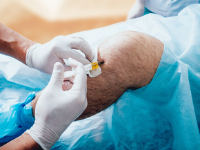
    Therapeutic Joint Injections

    Used to help with pain and limited motion, corticosteroid or visco supplement injections can provide relief and improved joint function.
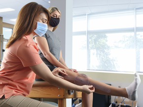
  • Physical Therapist Measuring The Range Of Motion Of A Patient's Knee

    Physical Therapy

    Our physical therapists can help you gain strength, increase range of motion, and restore mobility.

  • Tube of blood is placed in a medical centrifuge for plasma lifting

    Platelet-Rich Plasma

    This therapy involves injecting your own platelet-rich plasma into an arthritic joint or injured tendon. 

Surgical Approaches to Cartilage Repair and Preservation

  • Pen pointing a menisci on a plastic model

    Osteochondral Autograft Transplantation (OATS)

    Repairs damaged cartilage by harvesting and transplanting healthy cartilage from a non-weight bearing joint area.

  • Medically accurate 3D illustration of knee anatomy

    Matrix-Associated Autologous Chondrocyte Implantation (MACI)

    Biopsies of your healthy cartilage are used to grow additional cartilage in the lab for implantation.

  • MRI of knee joint

    Microfracture

    This technique makes small holes in the bone below the cartilage defect to increase blood supply and promote healing.

  • Doctor pointing on the knee problem point on x-ray film.

    Osteochondral Allograft (OCA)

    Donor bone and/or cartilage may be used to repair cartilage defects, with a low risk of reaction to the donor tissue.

  • Woman sitting on a park bench holding her knee

    Patellofemoral Arthroplasty

    Artificial joint resurfacing to treat patients with bone-on-bone osteoarthritis in the patellofemoral compartment of your knee.

  • Knee problem, x-ray view

    Meniscal Transplantation

    Typically appropriate for younger, active patients, this offers significant pain relief and improved function. 

  • Doctor pointing to area of hip joint x-ray

    Hip Cartilage Repair

    Removes unstable cartilage, while microfracture helps restore the joint with scar tissue that behaves like cartilage.

  • Doctor holding injured foot of patient

    Foot and Ankle Cartilage Repair

    Techniques include microfracture or drilling of the lesion, grafting cartilage and bone, or fixation of fragments with screws.

Xray image of knee joint

Your Treatment Plan and Recovery

First, your surgeon will obtain x-rays to evaluate your joint and determine whether or not you're a candidate for cartilage restoration surgery. Advanced imaging, such as an MRI, is usually necessary to assess cartilage defects and develop a treatment plan.

If your treatment plan involves surgery, you should know that most cartilage restoration surgeries are outpatient procedures, which means you'll go home after your procedure rather than stay in the hospital overnight. The majority of patients will begin slowly bending and straightening the joint soon after surgery to promote healing and improve motion.

Physical therapy typically begins a couple of weeks after surgery. You may be asked to wear a brace for the first 4-6 weeks after surgery; full recovery takes approximately 6-8 months for high-intensity sports or activities.

Meet Our St. Luke's Surgeons (2)

James Beckmann, MD

James T. Beckmann, MD

Orthopedic Surgery, Sports Medicine•Nampa, Meridian, Boise

Provider details
Justin Roth, DO

Justin S. Roth, DO

Orthopedic Surgery, Pediatric Orthopedics•Boise, Meridian

Provider details
St. Luke's Homepage

About St. Luke’s

Who We Are
News
Careers

Team Resources

For Providers
For Employees
For Contractors

Patient Resources

Financial Assistance
Mental Health Support
Medical Records
Patient Rights & Responsibilities

Essential Information

Accessibility
Price Transparency
En Español

Quick Links

Contact Us
St. Luke’s FitOne
St. Luke’s Health Partners
St. Luke’s Health Plan

DisclaimerNon-Discrimination/EEOPrivacy PracticesTerms of Use
© 2025 St. Luke’s Health System. All rights reserved.
FacebookLinkedinYoutubeInstagram